
The microspectrophotometer is a device that combines the optical capabilities of a microscope and a spectrophotometer. Microspectroscopy instruments are utilized for the purpose of measuring molecular spectra of samples that are microscopic in nature or microscopic features of samples that are on a large scale. These instruments are capable of measuring spectra from the deep ultraviolet (UV) to the near infrared (NIR) range. The microspectrophotometer has the capability to measure various properties of sub-micron-sized sample areas, including absorbance, reflectance, and emission spectra such as fluorescence, depending on its configuration. By incorporating specialized algorithms, the microspectrophotometer is capable of measuring the thickness of thin films and functioning as a colorimeter for microscopic samples.
What is microspectrophotometry?
Microspectrophotometry (MSP) is a scientific methodology employed to determine the absorption or transmission spectrum of a solid or liquid substance in either transmitted or reflected light. The technique is applicable for quantifying the emanation of electromagnetic radiation (luminescence, phosphorescence, fluorescence, or cathodoluminescence) from a specimen.
Microspectrophotometers are analytical instruments that integrate the magnification features of a microscope with the spectral analysis capabilities of a spectrophotometer. These devices are capable of obtaining transmission and reflectance spectra in the ultraviolet to near-infrared range of samples that are on a micron scale or smaller. Additionally, microspectrophotometers can be customized to measure fluorescence and luminescence spectra.
Why to use microspectrophotometer?
- The utilization of a microspectrophotometer enables researchers and professionals in the field of science and engineering to obtain spectra from minute sample regions in a non-invasive manner, without the need for direct physical contact with the sample.
- Various measurements can be conducted on a sample through different modes of light interaction, including transmission, reflection, scattering, and emission. This is exemplified in the image on the left, which displays OLED pixels.
- The UV-visible-NIR spectral range holds significant importance due to the higher absorption rate of various substances, including those that lack color, in the UV range compared to the visible and infrared regions. Hence, the utilization of a UV microscope spectrometer proves to be highly advantageous in the examination of various samples for diverse applications.
Microspectrophotometers are widely utilized across various disciplines and are present in both research laboratories and manufacturing plants.
- In industrial settings, they are employed for the purpose of ensuring quality control across a range of applications, including but not limited to color masks in flat panel displays and the thickness of films on semiconductor integrated circuits.
- Analytical laboratories employ microspectrometers to detect and measure minute samples, spanning from microfluidic kinetics to the comparison of fibers or paints by forensic chemists, the evaluation of gems or coal by geologists, the assessment of ink or paint color by process chemists, and the scrutiny of significant works of art by conservators. The microspectrometer is a versatile apparatus that can be employed in various fields.
How microspectrophotometer works?
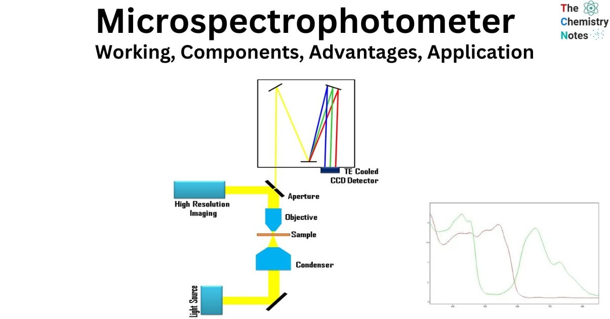
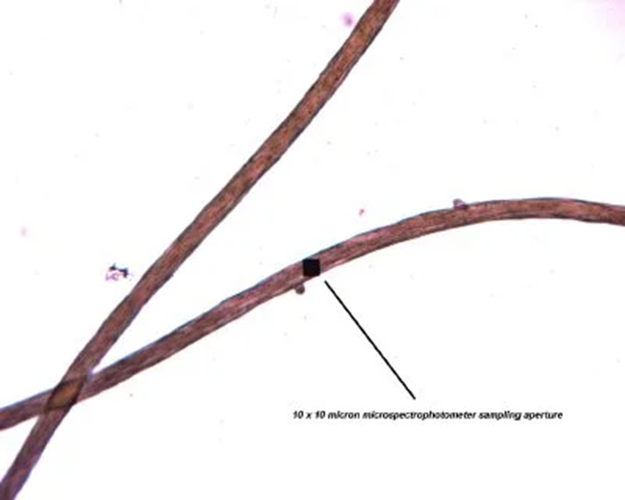
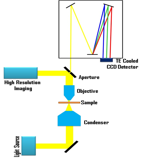
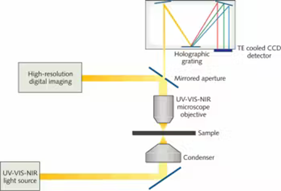
The operational mechanism of the microscope spectrometer and microspectrometer involves the emission of white light from a lamp on the microscope, which is subsequently directed towards the sample through a process of focusing. The preferential absorption of certain wavelengths of light by a sample is contingent upon the chemical structure and surrounding environment of the sample’s chromophore. The unabsorbed light is directed towards the entrance aperture of the spectrophotometer through the microspectrophotometer objective, where it is subsequently focused. Due to the mirroring of the aperture, a significant proportion of the incident light is directed towards a digital imaging system. The spectrophotometer aperture can be visualized superimposed on the sample, thereby facilitating the alignment of the system and acquisition of spectra. The visual representation depicted in Figure 1 illustrates the entrance aperture in the form of a black square. To obtain measurements, it is necessary to position the square over the sample by moving the stage.
Light that fails to reflect into the digital imaging system will traverse the aperture and enter the spectrophotometer. The optical grating is utilized to disperse the light into its constituent wavelengths, and the intensity of each wavelength is detected by a pixel on a Charge Coupled Device (CCD) detector. The information is stored by the computer, leading to the generation of an optical spectrum.
Various microspectroscopic methods are achieved through diverse illumination approaches. The selection of techniques is dependent upon the characteristics of the samples themselves. Opaque samples are typically illuminated using incident or reflectance illumination, while transparent samples are illuminated using transmitted light. The microspectrophotometer has the capability to be configured for the purpose of measuring various spectra, including transmission, absorbance, reflectance, and emission.
In the context of an operation, the process of obtaining a measurement is a simple and uncomplicated task. Initially, a dark scan is conducted to quantify the dark counts of the system. Subsequently, the spectrum resulting from a reference material is acquired. The spectral features of the reference material, light sources, optics, and CCD are encompassed within the reference spectrum. Subsequently, the spectrum emanating from the specimen is obtained and a computational method is employed to determine the corresponding spectra suitable for the given lighting scenario, specifically reflectance spectra in the case of measuring incident illumination. The computer automatically executes the algorithm and presents the outcome in the form of a spectrum.
Design and Components of microspectrophotometer
The instrument commonly referred to as the microspectrophotometer is recognized as a hybrid of an optical microscope and a spectrophotometer with exceptional sensitivity. The microspectrophotometer is a self-contained and custom-designed device, whereas the microscope spectrophotometer is an accessory that can be attached to a conventional microscope. The performance of add-on components is restricted by the inherent limitations of the microscope. An instrument that is integrated in nature circumvents the aforementioned limitations by being tailored toward the specific requirements of microspectroscopy.
- A microspectrophotometer is a device that combines a spectrophotometer with a custom-designed microscope.
- The microscope’s optical components and light sources exhibit exceptional quality and possess the capability to function across the ultraviolet, visible, and near-infrared spectra. This is in contrast to conventional microscopes, which are limited to operating within the visible range.
- The integration of a spectrophotometer and digital imaging system within the microscope enables the efficient capture of a high quantity of light from minute samples.
- Microspectrophotometer are highly versatile devices that can effectively determine the absorbance, transmittance, reflectance, and emission spectra (including fluorescence) of samples that are smaller than a micron.
Anatomy of microspectrophotometer
The current microspectrophotometer combines a spectrophotometer of high sensitivity with a microscope that is optimized for both spectroscopy and imaging.
- It is imperative that the microscope exhibits a functional spectral range spanning from the deep ultraviolet to the near-infrared, all the while ensuring optimal image and spectral fidelity.
- The utilization of standard microscopes is limited due to their optical configurations and light sources, which restrict their coverage of the visible spectrum.
- A personalized microscope is composed of lenses made of fused silica and other materials that are air-mounted, along with UV-enhanced mirrors. These components are arranged in a manner that ensures uniform illumination of a sample and produces a clear image across all spectral regions designated by the instrument.
- The provision of illumination is achieved through the utilization of modified xenon lamps, lasers, or a combination of the output from a deuterium lamp and a halogen lamp.
- The UV-VIS-NIR microscope functions similarly to a conventional compound microscope, thereby reducing the learning curve for users and facilitating the transition between various spectroscopic and imaging experimental setups.
In order to achieve accurate spectral outcomes, it is imperative that the spectrophotometer be designed specifically for microspectroscopy.
- A high dynamic range is necessary due to the frequent transition of users from transmission or reflectance spectroscopy to fluorescence spectroscopy while measuring a sample, in order to acquire diverse spectra from the identical location.
- The successful execution of this procedure necessitates the spectrophotometer to exhibit sensitivity while concurrently upholding a satisfactory level of spectral resolution. The stability of the microspectrophotometer poses a concern due to its single-beam nature, necessitating the acquisition of reference spectra prior to sample measurement.
- The central component of the system comprises a top-notch array detector that is equipped with thermoelectric cooling. The optimization of the monochromator’s optical design is aimed at achieving maximum light throughput, while simultaneously ensuring that the spectral resolution meets the customer’s acceptable standards.
The integration of the spectrophotometer with the microscope is a crucial aspect to consider. The utilization of calibrated variable apertures facilitates the selection of diverse sampling areas and enhances energy throughput, while simultaneously preserving spectral resolution and experimental reproducibility.
- The primary function of the microscope component is to provide illumination to the sample and direct the electromagnetic energy gathered from the sample into the spectrophotometer. To accomplish this task, it is imperative that the user possesses the ability to mentally construct a visual representation of the measurement region and also observe the adjacent sample.
- The spectrophotometer’s entrance aperture is situated at the identical focal plane as the sample image. A clear depiction of the aperture and sample fields is presented in a video image, which displays the aperture in precise focus over the sample and its surrounding field of view
- In the course of the operation, the sample stage is displaced until the image of the entrance aperture is aligned with the region of interest on the sample for measurement. Upon aligning the aperture with the sample region of interest, the spectrum is subsequently recorded. It is possible to store images of the measured sample while the aperture is in position.
- In numerous research applications, the alignment of the sample and aperture is performed through manual means. In the context of industrial operations, this process is frequently mechanized.
The arrangement of the microscope’s illumination schemes can vary depending on the type of experiment being conducted. Reflectance microspectroscopy can be performed from the deep UV to the NIR using white light, while fluorescence or phosphorescence microspectroscopy can be carried out by utilizing incident illumination with a white light source or a series of lasers mounted to the microspectrophotometer frame, along with a set of filters or a monochromator. The process of conducting transmission microspectroscopy involves the utilization of a white light that is directed onto the sample through the microscope’s condenser. In contrast to contemporary microscopes that possess a restricted spectral range of solely 450-700 nm, the microspectrophotometer microscope encompasses a broader range of 200-2200 nm.
Advantages of microspectrophotometer
There exist a multitude of benefits associated with the utilization of said tools.
- One notable benefit is the capability to obtain spectra from minute sample regions. Such instruments can evaluate sub-micron characteristics, necessitating minimal quantities of solid or liquid samples, and often without the need for extensive sample preparation.
- They can be employed for the purpose of cartography of the spectral properties of specimens with exceedingly fine spatial resolution.
- Microspectroscopic color comparisons are known to be more precise than those made by unaided human vision. This is due to the fact that microspectrophotometer are capable of analyzing spectra across a range of 200-2200 nm, which is beyond the range of human vision. These instruments are able to compensate for variations in lighting and measure the intensity of each wavelength of light.
- The microspectrophotometer has been developed to produce spectroscopic data of the utmost quality from minute samples, without any of the limitations associated with adapter modules employed in spectrophotometers designed for larger scales.
- Flexibility is an additional crucial characteristic.
- The UV-visible-NIR (UV-VIS-NIR) microspectrophotometer is capable of capturing both images and spectra through various modes of luminescence, such as absorbance, reflectance, and fluorescence, among others, using a single instrument.
- The microscope spectrometer offers a notable advantage in its capacity to employ apertures that enable precise control of the analysis area, thereby facilitating the quantitative comparison of solid specimens. The quantification of liquids can be accomplished through the utilization of flat capillaries or by means of a benchtop spectrophotometer equipped with a microcell. The latter enables the examination of samples with a volume as small as 10 μL. The utilization of a macro-spectrometer operating in the near-infrared region enables the examination of molecular overtone and combination bands, which finds application in diverse fields such as agriculture, medicine, and materials science.
Prior to the emergence of microspectroscopy, the sole method for examining numerous microscopic specimens entailed microchemical analysis followed by a form of visual inspection. Regrettably, these procedures result in the obliteration of a specimen, necessitate substantial quantities of a specimen, and are subject to the imprecisions inherent in the human visual perceptual system. UV-VIS-NIR micro spectrophotometers are capable of non-destructive analysis of microscopic samples and can identify sample variations that may not be discernible through visual observation. The factor of speed is a useful concern, as the duration of microchemical tests may range from a few minutes to several days for completion. The utilization of a microspectrophotometer enables the measurement of a spectrum within a short time frame of milliseconds.
Application of microspectrophotometer
- Spectroscopy of microscopic samples: The microspectrophotometer is a scientific apparatus utilized for the purpose of measuring the spectra of samples that are microscopic in nature. The utilization of spectrophotometers is widespread across various fields of study. In the semiconductor industry, engineers rely on spectrophotometers to determine the thickness of thin films. Similarly, forensic scientists employ spectrophotometers to analyze the dye present in individual textile fibers, as depicted on the left. Chemists also utilize spectrophotometers to measure the spectrum of nanocrystals.
- Forensic science: Microspectrophotometer are utilized in forensic science for the purpose of examining the absorbance and fluorescence spectra of various materials such as textile fibers, dyed hairs, paint chips, and colored glass fragments. In addition, they are utilized for the purpose of quantifying the reflectance and fluorescence spectra of documents, examining the non-destructive characteristics of ink and paper compositions, and scrutinizing handwriting details within a brief time frame. UV-absorbing compounds can be evaluated and compared using quartz optics by collecting spectra up to approximately 200 nm. Some samples have the ability to absorb ultraviolet radiation and subsequently emit these photons in the visible region of the electromagnetic spectrum. The luminescence that ensues, either fluorescence or phosphorescence, can subsequently be subjected to quantitative comparison by employing a range of excitation and barrier filters.
- Thickness of thin films: Microspectrophotometer are designed for the purpose of measuring the thickness of thin films utilized in the production of integrated circuits in semiconductor manufacturing. The microspectrophotometer is capable of obtaining reflectance or transmission spectra measurements, contingent on the transparency of the substrate’s small area being sampled. Analogous to the phenomenon of color alterations observed in oil floating on water, it is feasible to quantify these identical interference patterns by means of spectroscopic techniques. Subsequently, dedicated software is employed to compute the thickness of individual films that have been coated onto the substrate. Small-spot film thickness measurements continue to play a crucial role in quality control testing within the semiconductor industry. However, their application has expanded to encompass the development and production of various devices, including optics and flat-panel displays.
- Materials Science: The field of materials science is progressing due to the utilization of micro spectrophotometers, which possess the capability to measure transmission, reflectance, and emission spectra using a singular instrument. An area of significant advancement pertains to the phenomenon of surface plasmon resonance (SPR). The excitation of surface plasmons is achieved through the illumination of a metallic surface that is planar or consists of nanoscale metallic particles using light. Alterations in the optical properties of said materials manifest upon the interaction of said nanoparticles or surfaces with other materials. Microspectrophotometer are capable of obtaining reflectance and emission spectra of minute nanoparticle clusters, which can provide insights into the variations of the surface plasmon resonance (SPR) material’s spectra under different experimental conditions. This confers upon the researcher the capability to adjust the material to produce particular optical phenomena.
- This technique has the potential to be utilized in the analysis of metallo-enzymes, photosensitive proteins, or proteins containing co-factors, substrates, or products that exhibit UV-VIS absorption properties.
- The utilization of micro-spectrophotometry is viable in verifying the existence or non-existence of a substrate or co-factor, alongside the determination of the redox state of a protein. Additionally, it can facilitate the development of effective methods for initiating a reaction within the crystals or capturing transitional states.
- In the field of spectroscopy, the utilization of in-line micro-spectrophotometry has proven to be highly advantageous in the evaluation of radiation damage. This includes the identification and analysis of undesired redox reactions that may be initiated by the application of intense x-ray beams. Additionally, this technique is valuable in determining the optimal approach for data collection, with the aim of mitigating the effects of extensive radiation damage.
References
- Berglund, G.I., Carlsson, G.H., Smith, A.T., Szoke, H., Henriksen, A., and Hajdu, J. The catalytic pathway of horseradish peroxidase at high resolution. Nature 417, 463-468 (2002
- Pearson, A.R., Pahl, R., Kovaleva, E.G., Davidson, V.L., and Wilmot, C.M. Tracking X-ray-derived redox changes in crystals of a methylamine dehydrogenase/amicyanin complex using single-crystal UV/Vis microspectrophotometry. Journal of Synchrotron Radiation 14, 92
- https://biocars.uchicago.edu/facilities/micro-spectrophotometer/\
- https://www.laserfocusworld.com/test-measurement/test-measurement/article/16548076/spectrometers-microspectrophotometers-take-a-closer-look