
Microscopy is a method of making minute objects visible to the naked eye. It is utilized to examine cellular and subcellular components and for surface analysis. A microscope is an instrument, used to make small objects visible to the naked eye. A microscope is a device that enlarges objects causing them to appear larger.
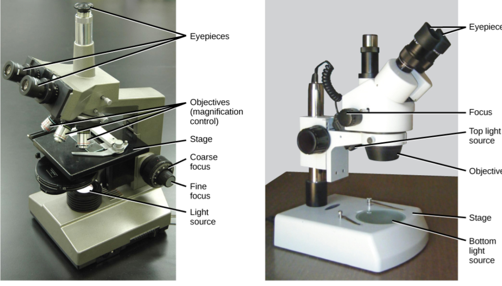
A simple microscope is a magnifying glass with a double convex lens and a short focal length. The compound microscope has a high resolution and two sets of lenses that produce a two-dimensional image of the sample.
Interesting Science Videos
Magnifying power and magnification
Magnification refers to how much larger an object appears when viewed via a microscope or a set of lenses within a microscope. Magnifying power of a compound microscope is defined as the ratio of the size of final image as seen through the microscope to the size of object seen with naked eye. In other words, it is the product of the magnification of the objective and magnifying power of the eyepiece.
Resolving power and resolution
The ability of a magnifying tool to differentiate two objects that are near together is referred to as resolving power. The limit of resolution and the resolving power are inversely related. The minimum distance between two points that enables their detection as two separate points, is known as the limit of resolution. As a result, the stronger the resolving power, the smaller the resolution limit.
Types of microscopy
There are mainly two types of microscope. They are:
- Light (optical) microscope
- Electron microscope
Light microscopy
Visible light is used as a source of illumination in a light microscope. Light microscopes have a resolution limit of roughly o.2 um and can magnify up to a maximum of approximately 1500 x. It is used to examine both living and dead specimens.
In this instrument light moves as a wave with crests and troughs. The light’s brightness is determined by the size of the crests and troughs. The frequency of a wave is the number of complete waves that occur in a unit of time, while the wavelength of a light wave is the separation between two successive crests. Light microscopes are further categories as
i. Brightfield microscope
ii. Darkfield microscope
iii. Phase -contrast microscope
iv. Flourescence microscopy
v. Confocal microscopy
Brightfield microscopy
It is the original and most widely used type of microscopy in which the specimen is viewed using transmitted light from the condenser lens.In a bright field microscope, light is directed at a condenser, then passes through the specimen, an objective lens, and finally to the eye through the eyepiece or ocular, which is a second magnifying lens. The object is visible through the light path.
Darkfield microscopy
Objects in a dark field microscope are irradiated from the side at a very low angle, making the background appear dark and the object visible against this dark background. It is a method for enhancing the contrast of translucent, unstained specimens.
Phase-contrast microscopy
It has a special type of condenser, objective and a special magnifier. Light passing from one
material into another of slightly different refractive index will undergo a change in the phase.
The refractive index of a live cell affects the phase of a light wave as it travels through it. The nucleus, for example, is relatively thick and dense and slows light traveling through it. As a result, its phase is altered in relation to light that has traveled through a nearby, thinner section of the cytoplasm.It is used to investigate living cells, which are often transparent to light.
Fluorescence microscopy
Certain chemical substances can absorb light and then partially reemit it as longer-wavelength light. Such materials are referred to be fluorescent, and the process is known as fluorescence. Only fluorescent light emitted by the sample is utilized in fluorescence microscopes to produce images. Most frequently, specific proteins or other compounds in cells and tissues are identified using fluorescence microscopy.
Confocal microscopy
Confocal means having a single point of focus. What this means for the microscope is that the focus of the final image matches or corresponds to the focus of the object. Lasers are the primary light sources in confocal microscopes. The microscope can filter out of focus information using confocal pinholes positioned in front of the image plane, which function as a spatial filter, allowing only the focused portion of the light to be imaged.
for detail information follow this link
https://pubmed.ncbi.nlm.nih.gov/12677057/
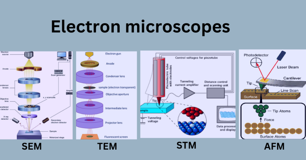
Electron microscopy
With one significant exception—the high-speed electron beams used in electron microscopy are focused using electromagnetic rather than optical lenses—the basic principles of electron microscopy are similar to those of light microscopy.
The electron microscope’s revolving power is extremely high because of the short wavelength of electrons. Living cells are not studied with electron microscopy. There are different types of electron microscopy. They are as below:
- Transmisssion electron microscope (TEM)
- Scanning electron microscope (SEM)
- Scanning tunneling electron microscope (STM)
- Atomic force microscopy
(Source: https://microbenotes.com/scanning-electron-microscope-sem/, https://emb-iitk.vlabs.ac.in/exp/transmission-electron-microscope/theory.html, https://www.ism.cnr.it/en/tempism/analysis/microscopy-diffraction-and-reflectometry/atomic-force-microscopy-afm.html, https://www.aps.org/publications/apsnews/200308/history.cfm)
Transmisssion electron microscope (TEM)
TEM is an imaging electron microscope in which an electron beam is passed through an ultra-thin sample specimen and an image is generated by the interaction of electrons transmitted from the sample specimen. It can give both image and diffraction data from a single sample surface. In 1993, Ernst Ruska and his research team created the first TEM with a resolving power greater than light. Germany installed the first TEM device for commercial use in 1939.
Scanning electron microscope (SEM)
Scanning electron microscope is a widely used imaging microscopy that gives high image resolution and unique image contrast. In this procedure, the sample is fixed and dried on the thin surface of heavy metal. Inside the microscope, a powerful electron beam rapidly scans the sample. Molecules in the sample are excited and produce secondary electrons, which are recorded by the detector and used to generate an image of the specimen. In addition to the topological information, SEM provides the element information as well as the compositional importance near to the surface region of the materials.
Scanning tunneling electron microscopy (STM)
STM evaluates surface topography using a tunneling current that is dependent on the distance between the metallic tip and the sample surface. When the tip and sample are linked to the voltage source, a small tunneling current flows between them. If the tip is close enough to the surface and the surface is electrically conductive, electrons will begin to leak across the gap between the probe and the sample. It is possible to measure this current. As tunneling is strongly dependent on the sample surface, any irregularity in size will reduce the rate of detection.
Atomic force microscopy (AFM)
AFM and scanning tunneling microscopy are comparable in that they both map the surface morphology of the material using an extremely sharp tip. However, AFM does not require tunneling electrons or tunneling currents. This technique fixes the tip to the end of a lengthy cantilever. The short tip’s interaction with the sample surface produces an interatomic force that bends the cantilever. While the tip is scanned over the sample surface, a detector detects the cantilever deflection. Using the measured cantilever deflection, the computer produces a surface topography map. AFM is used for the study of surfaces of insulators, semiconductors and electrical conductors also.
For further information:
https://www.slideshare.net/NithyaNandapal/electron-microscope-ppt
Applications of microscopy
A. In biological science
- The simplest use of microscopy is to examine a sample and determine its various components.
- For gram staining, is a method that provides the foundation for differentiating between Gram-positive and Gram-negative bacteria by staining the bacterial cell wall differently.
- Microscopic examination of bodily fluids:
- Light microscopy is used to count cells with a hemocytometer.
B. In helthcare
- Microscopy is essential in the research, formulation, and production processes of pharmaceutical product development.
- It helps to analyse blood and cellular features with remarkable clarity at higher and higher magnification.
- Use for pathogen detection.
C. For surface analysis
- It is used for the study of surfaces of insulators, semiconductors and electrical conductors.
- It helps to detrmine the element present in sample surface.
- It prvides topological,morphological, compositional and crystallographic information of the surfaces.
References
- J.Bhattrai, Introduction to electron microscopy for In-depth Surface Analysis, (1st edition), Balkhu Kathmandu Nepal.
- K. Pranav and M. Usha (2011). Biotechnology, A Problem Approach.Pathfinder Academy Private Limited, New Dehli.
- https://www.khanacademy.org/science/biology/structure-of-a-cell/introduction-to-cells/a/microscopy.
- Introduction to Microscopy.pdf.
- https://cattheni.edu.in/wp-content/uploads/2018/09/2.Microscopy-Principles-and-Types.pdf.
- https://uomustansiriyah.edu.iq/media/lectures/6/6_2018_12_17!11_51_16_PM.pdf.